

Infantile Scoliosis: Early Signs and Specialist Care
Infantile scoliosis is a three-dimensional spinal curve first recognized before age 3. It is uncommon and behaves differently from adolescent scoliosis: some idiopathic curves reduce during early growth, while others progress and may affect the developing chest and lungs.
A curve in a baby or toddler deserves pediatric assessment because early-onset scoliosis can be idiopathic or associated with congenital vertebral differences, neurological conditions, neuromuscular disorders, or syndromes. The goal is to identify the cause, understand the curve trajectory, protect development, and arrange the right specialist pathway.
This page is written for families worldwide, with additional relevance for Malaysia and Kuala Lumpur. It provides parent-friendly information without blaming normal caregiving or promising a particular outcome.
Infant-Specific Red Flags
A very young child cannot describe symptoms clearly. Changes in breathing, feeding, movement, development, or general health therefore deserve particular attention.
- Breathing difficulty, bluish color, recurrent respiratory distress, reduced exercise or play tolerance, or an unusual chest shape.
- Poor feeding, poor weight gain, persistent vomiting, marked fatigue, or failure to thrive.
- Weakness, unusual stiffness or floppiness, loss of developmental skills, unequal limb movement, or a new gait concern in a toddler.
- Bowel or bladder changes, abnormal reflexes, or another neurological concern.
- A rapidly increasing curve, marked trunk imbalance, or a chest that appears increasingly asymmetric.
- Persistent pain, unexplained distress, fever, systemic illness, or abnormal skin findings over the spine such as a deep dimple, hair patch, swelling, or discoloration.
These findings do not establish a specific diagnosis, but they justify prompt medical or pediatric specialist review.
What Infantile Scoliosis Means
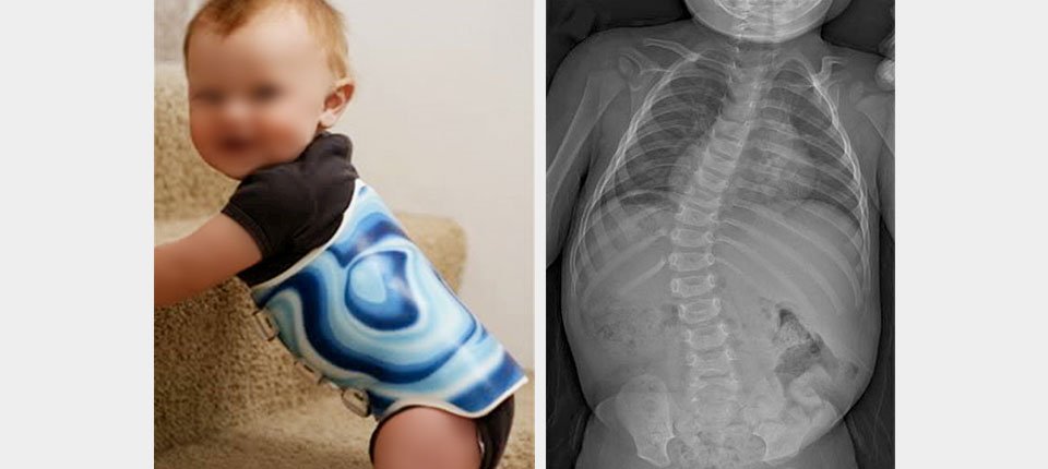 Infantile scoliosis is defined by age of onset, not by one cause. It usually refers to a structural curve first seen from birth to age 3. The curve may involve the thoracic, thoracolumbar, or lumbar spine and may show vertebral and rib-cage rotation as well as changes in the side profile.
Idiopathic infantile scoliosis means that no single cause is identified after appropriate evaluation. Other early curves may be congenital, neuromuscular, syndromic, or related to a neural-axis condition. The classification matters because natural history and treatment differ.
Resolving and Progressive Curve Patterns
Infantile idiopathic curves can follow very different paths. Repeat assessment is needed because one early measurement cannot identify the future with certainty.
| Feature | More reassuring pattern | Features raising progression concern |
|---|---|---|
| Curve behavior | The curve reduces or remains small on comparable follow-up. | The curve increases, becomes more rigid, or develops greater trunk and rib asymmetry. |
| Age and growth | Early mild asymmetry with favorable development and stable follow-up. | Very early structural deformity with substantial growth ahead and documented change. |
| Rib and vertebral relationship | Favorable rib phase and lower RVAD may support a resolving pattern. | Unfavorable rib phase or larger RVAD may add concern, but neither is an absolute predictor. |
| Cause and associated findings | No congenital, neurological, neuromuscular, or syndromic cause is identified. | Underlying vertebral, neural-axis, neuromuscular, syndromic, or thoracic abnormality is present. |
| Chest and respiratory development | Chest growth and breathing remain appropriate. | Thoracic restriction, recurrent respiratory concerns, or features of thoracic insufficiency appear. |
Causes and Conditions That Must Be Distinguished
Infantile scoliosis is a description of timing and curve pattern. The assessment must determine whether the curve is idiopathic or part of another condition.
- Idiopathic: no single cause is found. Genetic, developmental, growth, and molding-related influences have been studied, but ordinary carrying, holding, feeding, or sleeping choices are not established causes.
- Congenital: vertebrae form or segment differently before birth, such as a hemivertebra or unsegmented bar. Associated spinal cord, kidney, heart, or other organ findings may require coordinated investigation.
- Neural-axis: Chiari malformation, syringomyelia, tethered cord, or another spinal cord finding may be associated with an early curve.
- Neuromuscular: muscle tone, weakness, or neurological control changes the balance of forces on the growing spine.
- Syndromic or connective-tissue: the curve forms within a wider genetic or developmental condition.
- Functional or positioning-related asymmetry: some flexible postural findings change with position and must be distinguished from a persistent structural curve.
Signs Parents May Notice
Parents often notice asymmetry during bathing, dressing, carrying, tummy time, sitting, or early walking. A photograph or observation cannot diagnose scoliosis, but persistent change should be assessed.
- A visible curve or persistent leaning to one side.
- Unequal shoulder, rib, waist, or pelvic contours.
- One side of the chest appearing more prominent.
- An asymmetry that remains when the child changes position.
- Clothing or nappies appearing to sit unevenly.
- A developmental, neurological, breathing, feeding, or growth concern occurring with the spinal asymmetry.
How Infantile Scoliosis Is Assessed
Assessment is led by pediatric and spine specialists and is designed to define the curve, identify an underlying cause, estimate progression risk, and protect thoracic and neurological development.
Clinical and Developmental Review
History includes pregnancy and birth, development, feeding, breathing, growth, family history, associated conditions, and when the asymmetry was first noticed. Examination includes the spine, chest, skin, limb movement, tone, reflexes, hips, development, and cardiopulmonary status.
Standing, Sitting, or Supported Imaging
The imaging position is selected for the child’s age and ability. Front and side radiographs may document Cobb angle, curve location, rotation, sagittal shape, vertebral formation, rib relationships, and change over time. Radiation exposure and image comparability should be considered carefully.
RVAD and Rib Phase
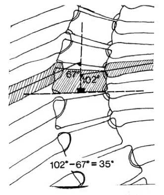
The rib-vertebra angle difference (RVAD) and rib phase were developed to help distinguish resolving from progressive infantile idiopathic curves. They remain useful descriptive tools, but later research shows that a threshold such as 20 degrees should not be treated as an individual guarantee. [1-3]
MRI and Other Medical Investigation
MRI may be considered to examine the spinal cord and neural axis, especially with a curve of meaningful size, atypical pattern, neurological finding, rapid progression, or another clinical concern. Studies of presumed idiopathic infantile and juvenile scoliosis have found clinically relevant neural-axis abnormalities in a subset of children. [4,5]
Multifactorial Infantile Progression-Risk Factors
These factors are interpreted together. Sanders and Risser stages are not valid maturity tools in infancy; age, developmental growth, curve behavior, rib findings, and the underlying diagnosis are more relevant.
| Category | Factors to assess | Why it matters |
|---|---|---|
| Age and development | Age at onset; developmental milestones; weight and height gain; thoracic growth; remaining years of growth. | The very young spine and chest have substantial growth ahead, creating both opportunity and risk. |
| Curve characteristics | Cobb angle; curve location and direction; rotation; flexibility; sagittal profile; rib phase; RVAD; vertebral formation. | The same Cobb angle can behave differently depending on pattern and structure. |
| Change over time | Comparable images; interval; progression or resolution; trunk and chest asymmetry; measurement variability. | Trajectory is more informative than one isolated image. |
| Cause and associated conditions | Idiopathic, congenital, neural-axis, neuromuscular, syndromic, chest-wall, or other causes. | The underlying diagnosis changes natural history and treatment. |
| Clinical findings | Breathing, feeding, growth, tone, strength, reflexes, development, pain or distress, skin findings, and systemic signs. | Medical and developmental findings can change urgency and investigation. |
| Family and care context | Ability to attend follow-up; anesthesia and casting burden; respiratory care; travel; home support; informed preferences. | The plan must protect safety, development, and family capacity over a long growth period. |
Structured Surveillance and Specialist-Led Care
The appropriate pathway is determined by the pediatric spine team after the cause, curve behavior, chest development, and overall health are understood. Immediate management may consist of structured surveillance, not necessarily immediate treatment.
Structured Active Surveillance
A mild curve with favorable features may be followed with defined intervals, comparable measurements, developmental review, and clear criteria for changing the plan. This is active surveillance, not indefinite waiting without a schedule.
Serial Casting
Serial casting is a specialist procedure used in selected progressive early-onset curves. Mehta’s work described using early growth as a corrective force, and subsequent literature supports casting as a meaningful option in selected infants. Casting requires specialist expertise, anesthesia planning, skin and respiratory monitoring, and repeated reassessment. [6,7]
Rigid Orthotic Bracing
Chiropractic Specialty Center® focuses on active, non-rigid, functional restoration protocols rather than rigid orthotic bracing. This page provides objective evidence to help families evaluate bracing claims.
A pediatric spine team may discuss an orthosis in selected situations. A prescribed brace should not be started, stopped, removed for imaging, worn for fewer hours, or materially changed solely because of website information. Any change must be coordinated with the prescribing practitioner and orthotist.
Growth-Friendly Procedures and Surgery
When a progressive curve threatens spinal, thoracic, neurological, or overall development, a pediatric spine surgeon may discuss growth-friendly instrumentation or another operation. The decision is individualized and should address the purpose, expected growth, number of procedures, implant and anesthesia burden, infection and neurological risks, alternatives, and the family’s informed preferences.
Registered Physiotherapy and CSC Coordination
Infants cannot perform active self-correction in the way an older child can. Registered physiotherapy may nevertheless support age-appropriate movement, respiratory development, positioning, function, and family guidance when coordinated within the pediatric plan. CSC’s immediate contribution is careful recognition, parent education, and referral coordination; active curve-specific rehabilitation becomes increasingly relevant as the child develops the ability to participate.
Thoracic Growth and Breathing
 Early-onset scoliosis can affect more than the spine because the ribs, chest, and lungs are still developing. Thoracic insufficiency syndrome describes an inability of the thorax to support normal breathing or lung growth. It is associated particularly with severe curves, fused ribs, congenital differences, and restricted chest development. [8]
Breathing difficulty, recurrent respiratory illness, poor growth, or a worsening chest deformity should prompt pediatric medical and spine review. Protecting thoracic development is one reason progressive infantile scoliosis is managed differently from an adolescent curve.
Practical Guidance for Families
- Do not blame ordinary holding, carrying, sleeping, or feeding practices for an idiopathic curve.
- Keep scheduled pediatric and imaging reviews even when the child appears comfortable.
- Bring previous images and reports so change can be compared accurately.
- Record developmental, feeding, breathing, skin, movement, and comfort changes between visits.
- Ask how each measurement will change the plan rather than focusing on one number alone.
- Seek an independent pediatric spine opinion when a major procedure is proposed or uncertainty remains.
Frequently Asked Questions About Infantile Scoliosis
The questions below cover what people most often want clarified about Infantile Scoliosis | Early Detection & Non-Surgical Care.
1. What age is infantile scoliosis diagnosed?
Infantile scoliosis is first recognized before age 3. It is part of early-onset scoliosis, a broader group that includes curves appearing before age 10. A curve in a baby or toddler should be assessed in an age-appropriate pediatric pathway.
2. Can infantile scoliosis resolve as a child grows?
Some mild idiopathic infantile curves reduce or resolve during early growth, while others progress. The direction cannot be predicted safely from one Cobb angle alone. Age, curve pattern, rib phase, RVAD, underlying cause, development, and repeat imaging are considered together.
3. What is the rib-vertebra angle difference, or RVAD?
RVAD is measured on a front-view spinal radiograph by comparing the rib-vertebra angles on the two sides near the curve apex. It can contribute to prognosis, but a cutoff such as 20 degrees is not an individual guarantee and should be interpreted with the whole clinical and imaging picture.
4. Why might MRI be considered in infantile scoliosis?
MRI can help identify neural-axis findings such as syringomyelia, Chiari malformation, tethered cord, or another condition that may accompany an early-onset curve. The pediatric specialist decides whether MRI is appropriate based on curve size, pattern, neurological findings, age, and other clinical features.
5. What signs may suggest infantile scoliosis?
Parents may notice a persistent curve, uneven shoulders or trunk, a rib prominence, one side of the chest appearing different, a preferred leaning position, or clothing that fits asymmetrically. These observations do not confirm scoliosis, but they justify pediatric assessment.
6. How is progressive infantile scoliosis managed?
Management is led by a pediatric spine team and may include structured surveillance, serial casting, selected orthotic strategies, respiratory or neurological evaluation, and growth-friendly surgical procedures when necessary. The pathway depends on the cause, curve behavior, thoracic development, and the child’s overall health.
7. When does an infant with a spinal curve need urgent review?
Prompt medical review is appropriate for breathing difficulty, poor feeding or poor growth, weakness or loss of developmental skills, bowel or bladder changes, marked pain or distress, a rapidly increasing curve, abnormal skin findings over the spine, or other neurological or systemic concerns.
Related Scoliosis Guides
- Types of scoliosis by age and cause
- Juvenile scoliosis
- What scoliosis means in three dimensions
- Scoliosis examination and assessment
- Scoliosis surgery considerations
- Scoliosis care options
Clinical Information and Referral in Kuala Lumpur
Infantile scoliosis requires pediatric medical and spine-specialist leadership. For general scoliosis education, referral coordination, and current CSC contact information, use the approved pages below. Urgent pediatric symptoms should be directed to appropriate medical care without delay.
- Primary scoliosis clinical information
- Bukit Damansara center information
- Bandar Sri Damansara center information
- Current contact and referral information
Core Academic References
- Scott JC, Morgan TH. The natural history and prognosis of infantile idiopathic scoliosis. Journal of Bone and Joint Surgery British Volume. 1955;37-B(3):400-413.
- Mehta MH. The rib-vertebra angle in the early diagnosis between resolving and progressive infantile scoliosis. Journal of Bone and Joint Surgery British Volume. 1972;54-B(2):230-243.
- Wynne-Davies R. Infantile idiopathic scoliosis: causative factors, particularly in the first six months of life. Journal of Bone and Joint Surgery British Volume. 1975;57-B(2):138-141.
- Gupta P, Lenke LG, Bridwell KH. Incidence of neural axis abnormalities in infantile and juvenile patients with spinal deformity. Spine. 1998;23(2):206-210.
- Zhang W, Sha S, Xu L, et al. The prevalence of intraspinal anomalies in infantile and juvenile patients with presumed idiopathic scoliosis: a MRI-based analysis of 504 patients. BMC Musculoskeletal Disorders. 2016;17:189.
- Mehta MH. Growth as a corrective force in the early treatment of progressive infantile scoliosis. Journal of Bone and Joint Surgery British Volume. 2005;87-B(9):1237-1247.
- Alassaf N, Tabard-Fougere A, Dayer R. Casting in infantile idiopathic scoliosis as a temporising measure: a systematic review and meta-analysis. SAGE Open Medicine. 2020;8:2050312120925339.
- Campbell RM Jr, Smith MD, Mayes TC, et al. The characteristics of thoracic insufficiency syndrome associated with fused ribs and congenital scoliosis. Journal of Bone and Joint Surgery American Volume. 2004;86(2):399-408.
Author and Publisher
Authored by Yama Zafer, D.C., Doctor of Chiropractic (Cleveland University-Kansas City), registered with the Malaysian Traditional and Complementary Medicine Council in the recognized practice area of chiropractic. He is the founder and director of Chiropractic Specialty Center® and has more than 30 years of clinical experience. Physiotherapy services are delivered by registered physiotherapists.
skoliosis.my is the scoliosis education website published by Chiropractic Specialty Center®.
Last Updated
Infantile Scoliosis: Early Signs and Specialist Care was last updated on August 24, 2026.